名称 : 460MPa抗震耐蚀耐火钢高温保持15minEBSD
英文名称 : 460MPa anti-seismic and corrosion resistant refractory steel keeps 15minEBSD at high temperature
材料 : 533
委托单位 : 钢铁研究总院工程用钢所
实验单位 : 钢研纳克材料检测股份有限公司
实验方法 : EBSD分析
实验设备 : FEI Quanta650热场发射扫描电子显微镜
实验条件 : 高温保持15分钟
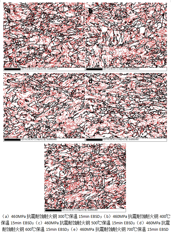
说明 : 在本研究中,将水磨抛光后的试样,在10 ml HClO4 + 90 ml C2H5OH混合溶液中进行电解抛光,电压与时间分别为10~20 V和10~15 s。使用配备有Oxford Nordlys F+型EBSD附件的FEI Quanta 650热场发射电子显微镜进行晶体学数据采集,并获取界面取向差分布等,扫描面积为80×100 μm,扫描步长为0.1~0.2 μm,观察发现在300℃到600℃保温15min的样品中,贝氏体组织中小角度界面逐渐降低,直到在700℃保温15min样品中,观察到贝氏体组织中小角度界面再次增多。
英文说明 : In this study, water polished samples were electrolytically polished in 10 mL HClO4 + 90 mL C2H5OH mixed solution at a voltage of 10~20 V and 10~15 s, respectively. The FEI Quanta 650 thermal field emission electron microscope equipped with Oxford Nordlys F+ EBSD attachment was used to collect crystallography data and obtain the interface orientation difference distribution. The scanning area was 80×100 μm and the scanning step size was 0.1~0.2 μm. It was observed that the small and medium Angle interface of bainite tissue decreased gradually in the sample held at 300℃ to 600℃ for 15min, until the small and medium Angle interface of bainite tissue increased again in the sample held at 700℃ for 15min.
数据来源 : EBSD分析
重点项目名称 : 建筑结构用抗震耐蚀耐火钢-新一代建筑结构用钢的组织调控和性能稳定化关键技术研究数据集