名称 : DZ2(模铸)钢边部SEM组织
英文名称 : SEM structure of DZ2 (die cast) steel edge
材料 : 876
委托单位 : 钢铁研究总院
实验单位 : 钢铁研究总院中心实验室
实验方法 : 扫描电镜观测
实验设备 : FEI Quanta650热场发射扫描电子显微镜
实验条件 : 室温
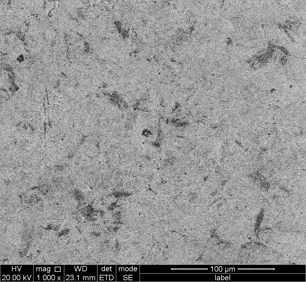
说明 : 在高速车轴DZ2(模铸)钢的边部切取10×10×8的镜像样品,在国产UltrMet6型预磨机上进行预磨,然后在150、320、600和1000号砂纸上依次水磨后,在PG-2C型抛光机上进行机械抛光,抛光后的试样置于4 ml 硝酸(HNO3) + 96 ml 无水乙醇(CH3CH2OH)混合溶液浸蚀10~15 s,采用FEI Quanta650热场发射扫描电子显微镜分析,如图所示,由图可知试样中组织基本为回火马氏体,在图中可以发现有少量的贝氏体组织。
英文说明 : A mirror sample of 10×10×8 was cut from the edge of high-speed axle DZ2 (die cast) steel, which was pre-ground on domestic UltrMet6 pre-grinding machine, and then water ground on sandpaper 150, 320, 600 and 1000 in turn, and then mechanically polished on PG-2C polishing machine. After polishing, the sample was immersed in a mixed solution of 4 mL nitric acid (HNO3) + 96 ml anhydrous ethanol (CH3CH2OH) for 10~15 s. FEI Quanta650 thermal field emission scanning electron microscope was used for analysis, as shown in the figure. It can be seen from the figure that the tissue in the sample is basically tempered martensite. A small amount of bainite tissue can be seen in the figure.
数据来源 : 检测数据
重点项目名称 : 苛刻环境下铁路车辆关键部件用钢-高速车轴钢基础研究数据集